
Basic Eye Anatomy South Bay Ophthalmology
Apr. 29, 2023 To understand the diseases and conditions that can affect the eye, it helps to understand basic eye anatomy. Here is a tour of the eye starting from the outside, going in through the front and working to the back. Eye Anatomy: Parts of the Eye Outside the Eyeball The eye sits in a protective bony socket called the orbit.
Anatomy of the Eye Human Eye Anatomy Owlcation
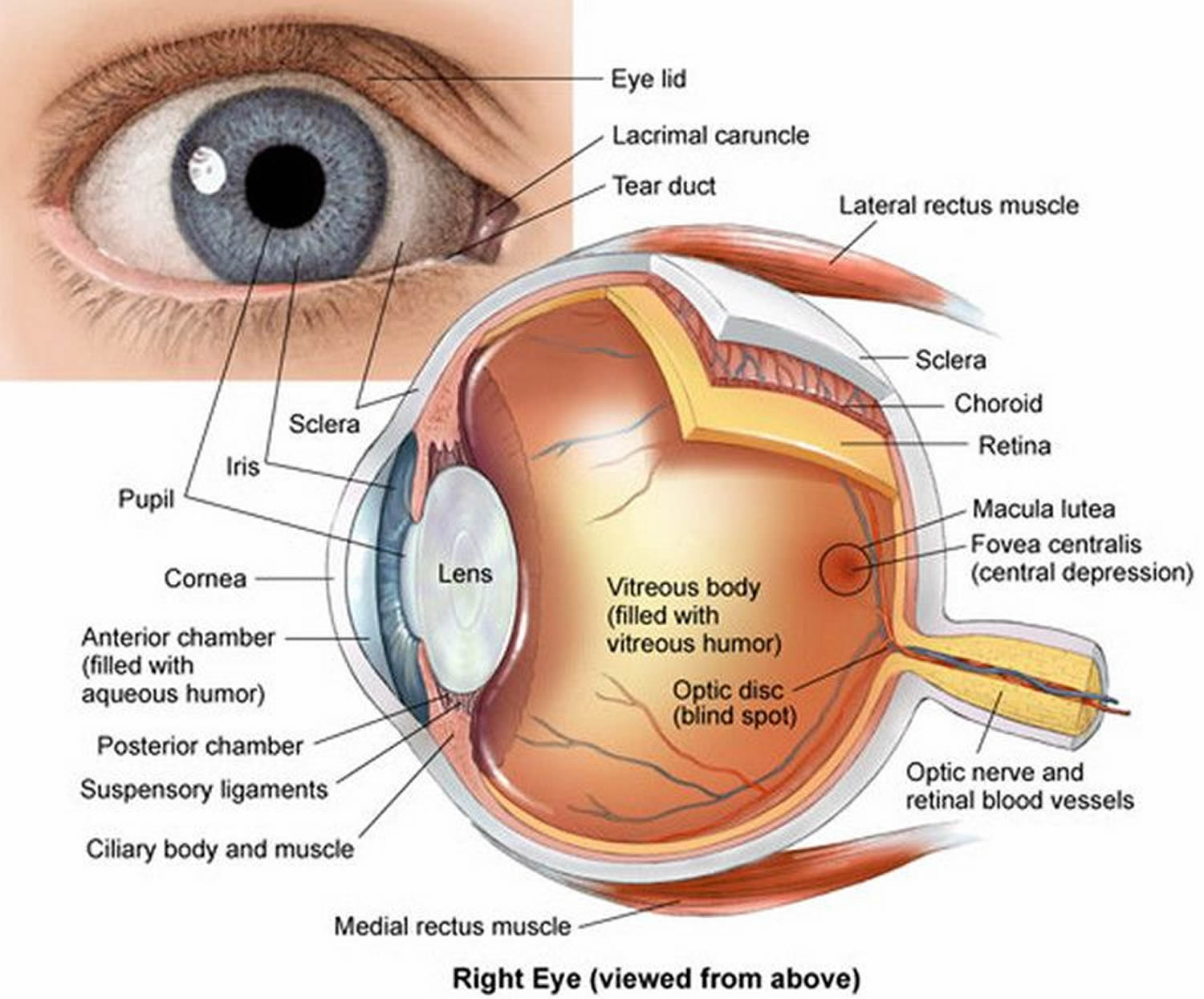
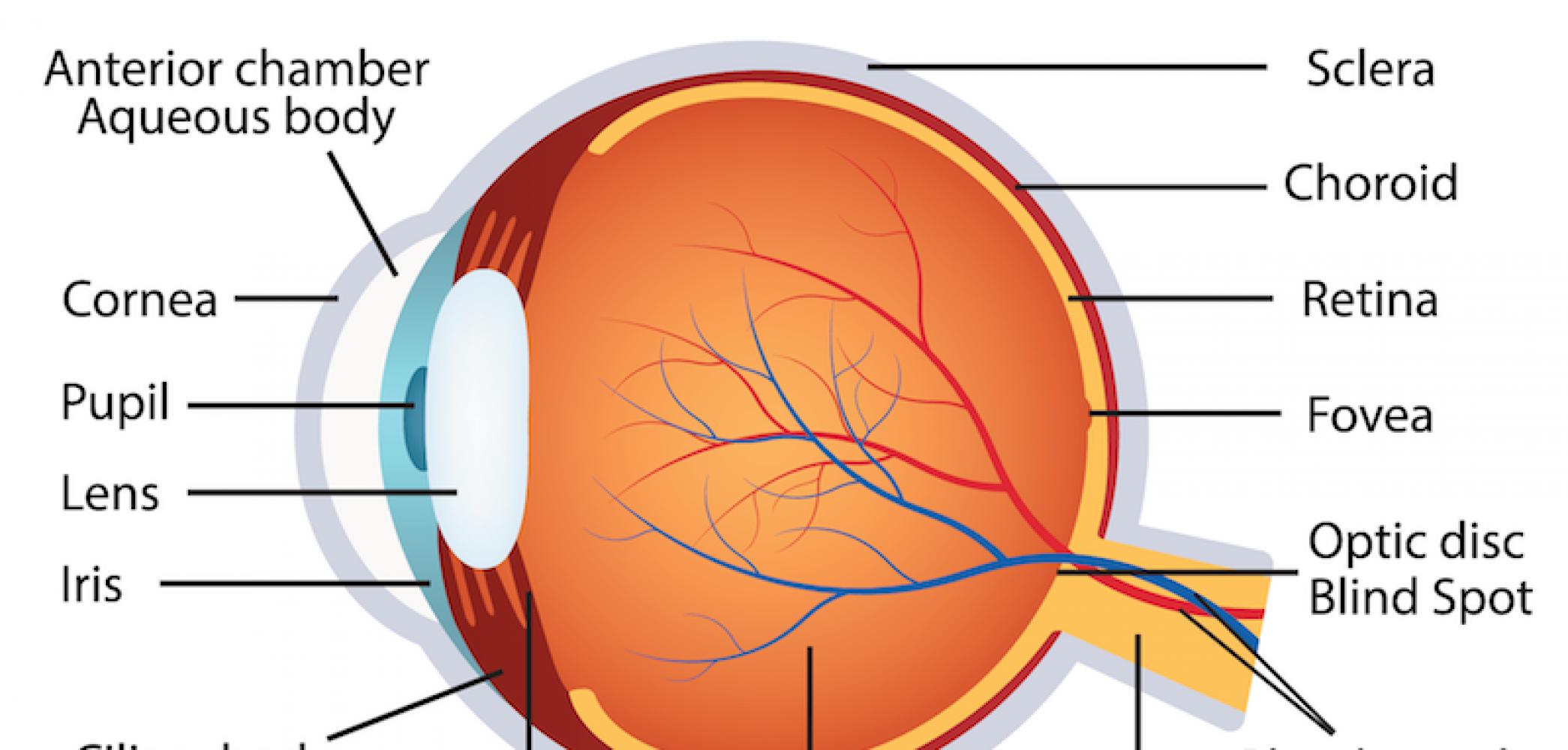
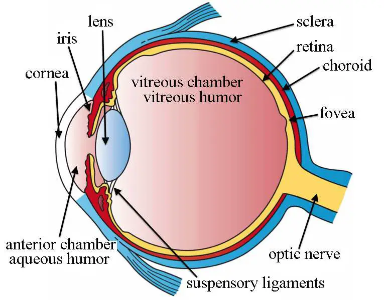
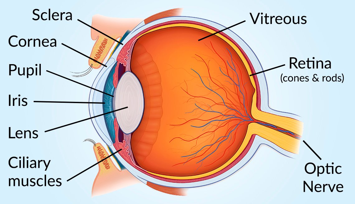
The main parts of the human eye are the cornea, iris, pupil, aqueous humor, lens, vitreous humor, retina, and optic nerve. Light enters the eye by passing through the transparent cornea and aqueous humor. The iris controls the size of the pupil, which is the opening that allows light to enter the lens. Light is focused by the lens and goes.
How to draw human eye diagram for beginners YouTube
Diagram of the Eye Posted in Eye Health, Uncategorized | August 5, 2018 Even though the eye is small, only about 1 inch in diameter, it serves a very important function - the sense of sight.
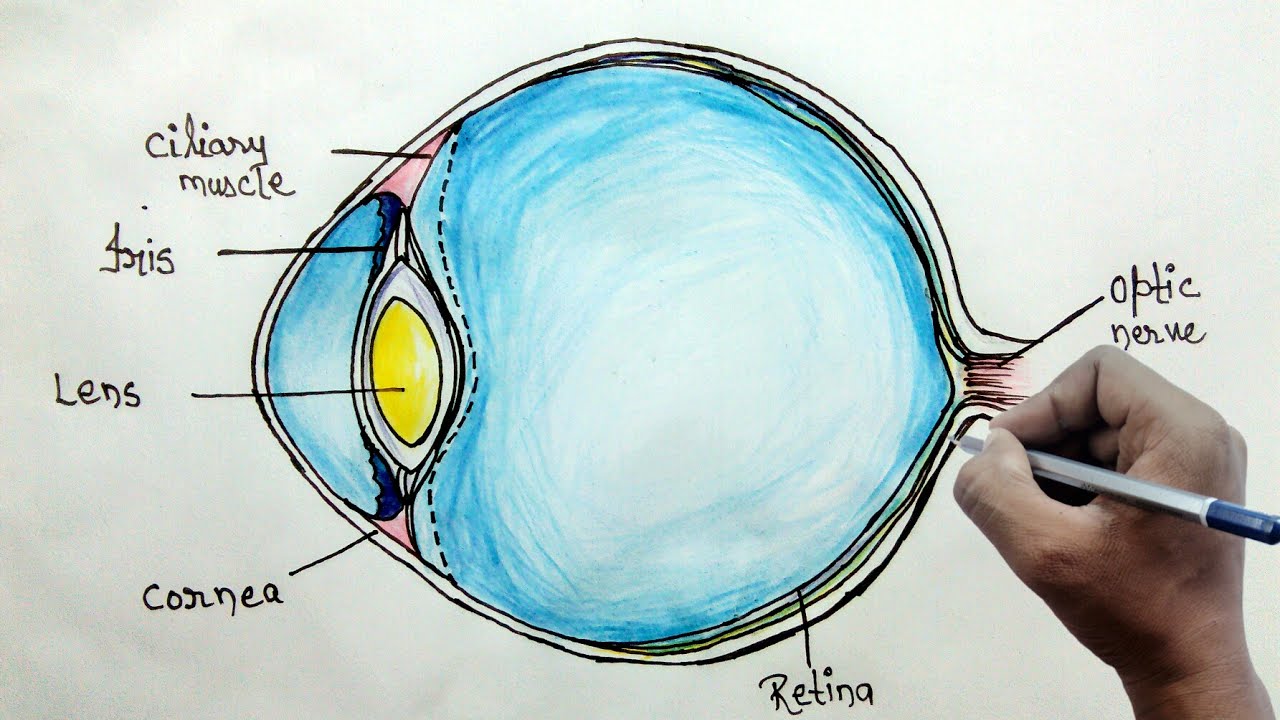
Eye Diagram drawing CBSE easy way draw Human eye anatomy Step by step for beginners
Lens - This focuses light onto the retina. Retina - Light-sensitive layer at the back of the eye. It is made up of rods and cones. Rods - Sense cells that help us see the shapes of things. Cones.

OUR EYES WORK LIKE CAMERA’S! Discovery Eye Foundation
Overview of the Eyes VIDEO Each photoreceptor is linked to a nerve fiber. The nerve fibers from the photoreceptors are bundled together to form the optic nerve. The optic disk, the first part of the optic nerve, is at the back of the eye.
HUMAN EYE (STRUCTURE, IMAGE FORMATION AND DIFFERENCE BETWEEN RODS AND CONES) « SimpleBiology
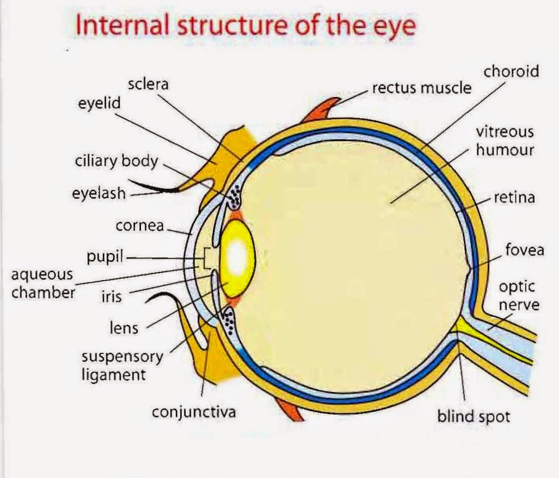
Eyelid anatomy The eyelids are soft tissue structures that cover and protect the anterior surface of the eyeball. The anatomy of the eyelid may seem complex, but if we dissolve its multi-layered structure it is actually quite simple: Skin
Human Eye Anatomy Parts of the Eye and Structure of the Human Eye
How to learn the parts of the eye. Found within two cavities in the skull known as the orbits, the eyes are surrounded by several supporting structures including muscles, vessels, and nerves. There are 7 bones of the orbit, two groups of muscles (intrinsic ocular and extraocular), three layers to the eyeball. and that's just the beginning.

Diagram showing the different parts of the eye Parts of the eye, Eye health, Free homeschool
Light is focused primarily by the cornea - the clear front surface of the eye, which acts like a camera lens. The iris (colored part) of the eye functions like the diaphragm of a camera, controlling the amount of light reaching the retina by automatically adjusting the size of the pupil (aperture). The eye's crystalline lens is located.
:max_bytes(150000):strip_icc()/eye-conjunctiva-871453538-5a26c6ad22fa3a0037d5edad.jpg)
How the Human Eye Works (Structure and Function)
6 min read Your eye is a slightly asymmetrical globe, about an inch in diameter. The front part (what you see in the mirror) includes: Iris: the colored part Cornea: a clear dome over the iris.
humaneyeanatomy La Pine Eyecare Clinic
A human eye is roughly 2.3 cm in diameter and is almost a spherical ball filled with some fluid. It consists of the following parts: Sclera: It is the outer covering, a protective tough white layer called the sclera (white part of the eye). Cornea: The front transparent part of the sclera is called the cornea.
Simple eye diagram
Anatomy of the Human Eye. Eyes are one of the most important organs of the body. A healthy pair of eyes means a clear vision, which plays a major role in day-to-day life and quality of experiences.

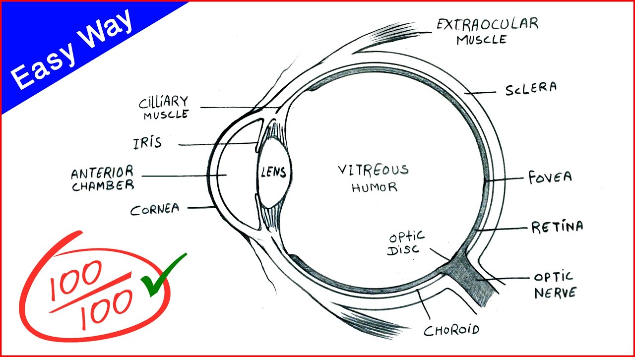
Simple Diagram Of Human Eye With Labelling Human Eye Diagram Class 10 How To Draw Human Eye
The iris is a flat, thin, ring-shaped structure sticking into the anterior chamber. This is the part that identifies a person's eye colour. The iris contains both circular muscles going around the pupil and radial muscles radiating toward the pupil. When the circular muscles contract, they make the pupil smaller.
Vision and Eye Diagram How We See
The human eye is a sensory organ,. One basic model describing the geometry of the optical system is the Arizona Eye Model.. Schematic diagram of the human eye. It shows a horizontal section through the right eye. The eye is made up of three coats, or layers, enclosing various anatomical structures..

Anatomy of the Eye Human eye diagram, Eye anatomy diagram, Eye anatomy
How Do the Eyes Work? Eye Anatomy (16 Parts of the Eye & What They Do) Summary How Do the Eyes Work? Light is reflected when you focus on an object and enters the eye through the cornea. As the light passes through, the dome-shaped nature of the cornea bends light, enabling the eye to focus on fine details.
/GettyImages-695204442-b9320f82932c49bcac765167b95f4af6.jpg)
Structure and Function of the Human Eye
Iris: regulates the amount of light that enters your eye. It forms the coloured, visible part of your eye in front of the lens. Light enters through a central opening called the pupil. Pupil: the circular opening in the centre of the iris through which light passes into the lens of the eye. The iris controls widening and narrowing (dilation and.
/GettyImages-1128675065-e4bac15b0f39449dba31f25f1020bc8a.jpg)
An Overview of Eye Anatomy
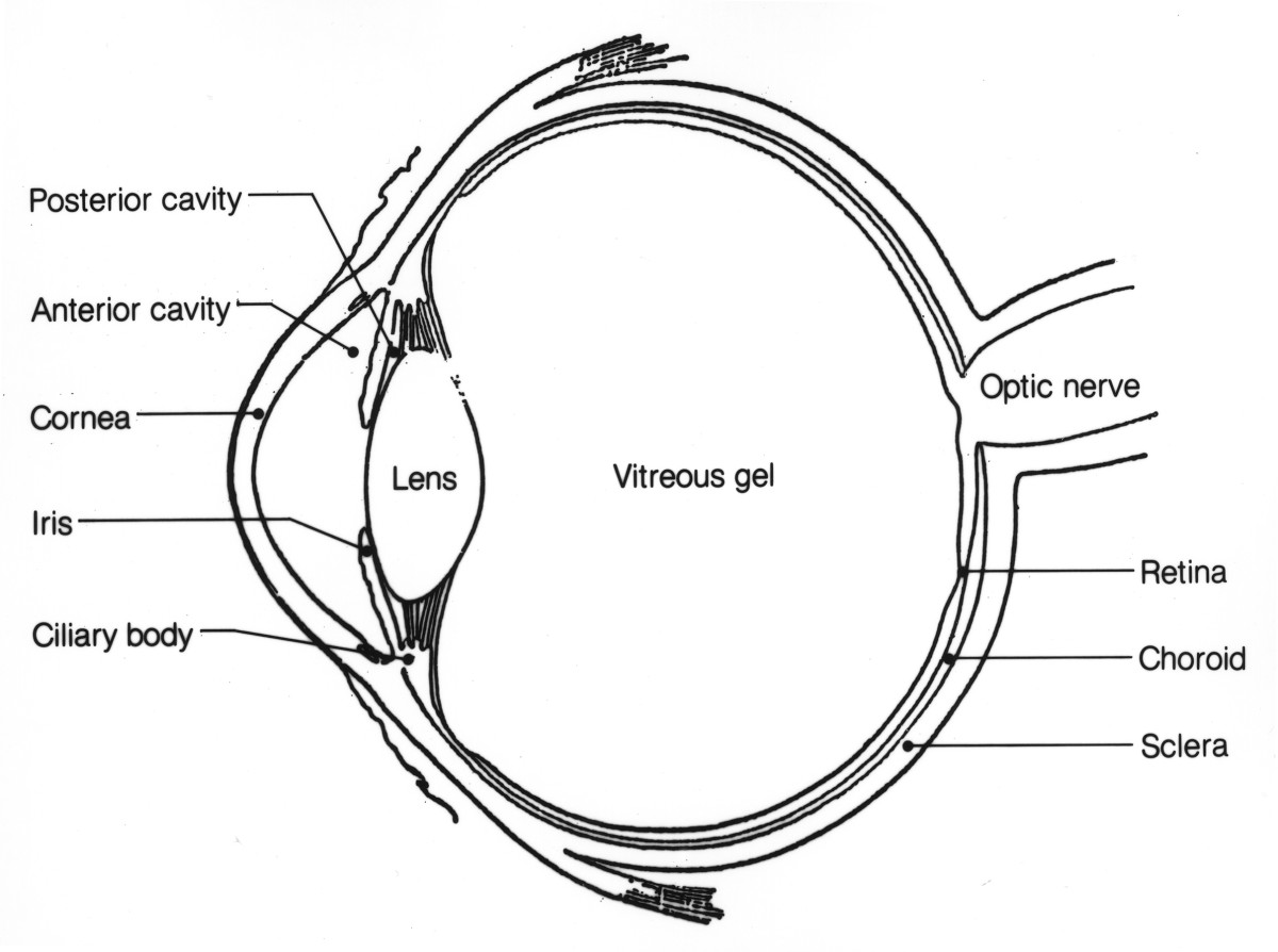
Choroid. The thin, blood-rich membrane that lies between the retina and the sclera and is responsible for supplying blood to the outer portion of the retina. Ciliary body. The part of the eye that produces aqueous humor. Cornea. The clear, dome-shaped surface that covers the front of the eye. Iris. The colored part of the eye.